Once upon a time, many of the world’s creatures had, in addition to the pair of eyes we are accustomed to seeing in animals, a third eye, perched on their head. A vestige of an early and mysterious stage of evolution, this eye still exists, in various forms, in several species of vertebrates, and even humans have, hidden deep in the brain, a special organ, still too little known, which was once associated with this eye.
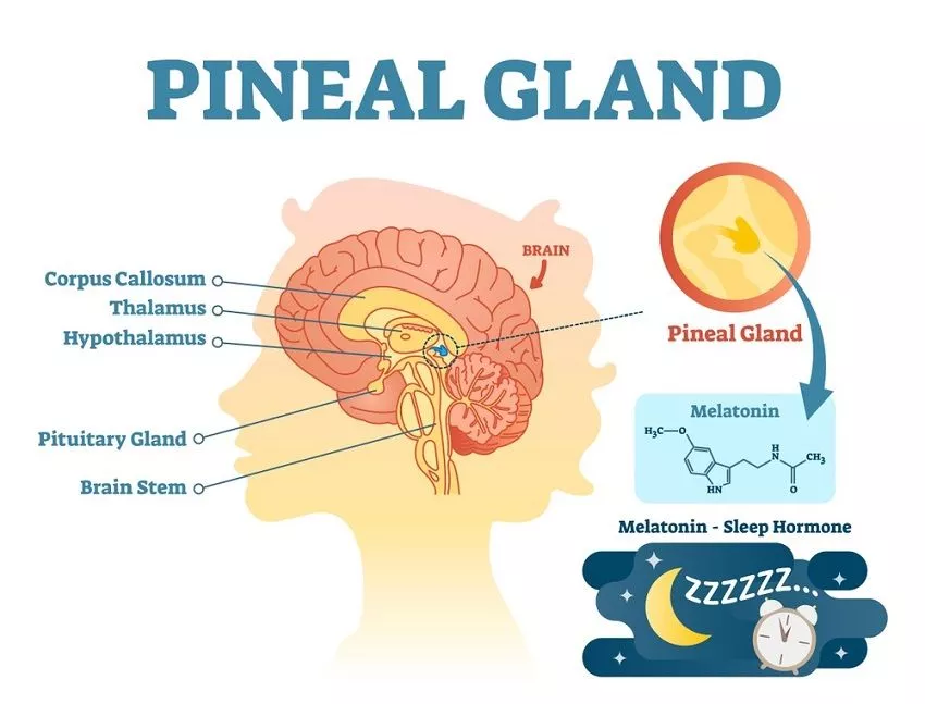
In humans, it’s the pineal gland, or epiphysis, a small organ located almost in the center of the brain and whose secretory function is still too little understood. It is known that it is influenced by light, that it is related to the day-night cycle, sleep and sexual development, but we still have much to learn about it.
But how did we get on with the pineal gland and how can its mysterious connection to light and darkness be explained?
During the evolution of vertebrates, they were endowed by nature with a so-called parietal eye (or pineal eye), which, together with the pineal gland, formed a functional whole that forms part of the epithalamus, a region of the brain. Over time, the most evolved of vertebrates – birds and mammals – have lost this third eye, the parietal eye, retaining only the pineal gland. Other vertebrates, however, still have vestiges of this photosensitive organ, and in some its structure is disturbingly similar to that of ordinary eyes.
A number of primitive vertebrates – placoderm fish, ostracoderms, crossopterygians, even some early tetrapods – have left us fossils that have, in the skull, a depression (like an orbit) that seems to have housed a parietal organ. In some of the skulls there is also a foramen (an opening) in the scalp, through which the parietal eye receives light. But does it still exist in today’s vertebrates?
Given that it’s an ancient feature of vertebrates, which tends to disappear as they have evolved into more modern forms, it is logical that if it still exists in some species today, then we should look for it in old species, members of the groups that emerged early. And, indeed, the parietal organ is found in some amphibians and in some primitive species of reptiles and fish.
In most of them, it is reduced; its contour can still be seen on the dorsal surface of the head and it is possible that it still has a role in photoreception, but we do not know much about it. In others, however, it has retained its sensitivity to light and has a structure similar to that of the lateral eyes. Not coincidentally, such a situation is found in very old, surviving species of distant eras, those we often call living fossils.
Cyclostomies are ancient fish, which appeared early in evolution; in fact, these creatures are not even considered by all zoologists as fish, but as a separate group of vertebrates, the most primitive of vertebrates. But, for simplicity, let’s adopt the concept of those who call them fish. They are creatures with a long, cylindrical body, like eels, but with a cartilaginous skeleton, having other primitive characters that place them at the base of the phylogenetic tree of vertebrates. Their defining peculiarity is the lack of jaws (hence the older name given to the group, Agnatha – “jawless fish”).
Their mouth is circular, resembling a suction cup with several rows of keratinous teeth; with its help, the parasitic species of this group (not all of them are parasitic) catch themselves on the bodies of other fish, feeding on their blood. In some species in this group, the presence of photosensitive parietal organs has been found, which most researchers believe would have a role in regulating the behavior of fish depending on the rhythm of day-night and season.
In his book “The Third Eye”, zoologist Richard Marshall Eakin (1910-1999) mentions that in the cyclostomies studied, two interconnected formations were discovered – which he calls the pineal eye and the parietal eye – in which there are cells with photoreceptor structure, as microscopic research has shown.
Two other animals – this time reptiles – in which the parietal eye has been remarkably preserved are the two species of tuatara, New Zealand reptiles that “date” from the time of the dinosaurs. They have perished, but the small tuatara have survived the times, surviving to this day, although their situation in the contemporary world is not one of the most prosperous.
The two species of tuatara, Sphenodon punctatus and Sphenodon guntheri, are the only survivors of an ancient group called Rhynchocephalia, which appeared about ca. 200 million years ago. The parietal eye of the tuatars is among the best studied and, from what we know so far, these species have the most complete and best preserved (evolutionarily speaking) parietal eye, of all the existing tetrapods.
Research shows that it has a complex structure, with cornea, lens and retina made up of photoreceptor cells, even if, in some aspects, its composition is more reminiscent of the eye of octopuses than that of vertebrates, say researchers I. R. Schwab and iG. R. O’Connor, of the University of California, in a paper published in 2005.
In newly hatched chicks, the cornea appears as a “patch” of transparent membrane on the top of the head; after a few months, it will be covered with opaque scales on the skin. He still remains able to perceive light, some scientists believe (who, however, are still quite confused when it comes to explaining the function of this mysterious “third eye” of reptiles).
They talk about the existence of a “pineal complex” in the tuatara, made up of two components: the third eye (the parietal eye) and the pineal gland. Most of the specialists who studied believe that the pineal eye would be a kind of solar “dosimeter”, with the role of estimating the amount of light, based on which it determines what time of day it is, what season, and depending on these “findings” the reptile’s body reacts most appropriately.
Behavioral changes depending on the circadian and seasonal rhythm are coordinated by the pineal gland, functionally connected to the parietal eye; it secretes, in reptiles, the hormone called melatonin, which intervenes in establishing the alternation of sleep and wakefulness and in thermoregulation.
In any case, the presence of the parietal eye, in different stages of evolution, is a proof that it once had an important role in the life of animals, a role then taken over by the two lateral eyes of vertebrates. The parietal eye is considered by many researchers as a vestigial organ, a remnant of an anatomical structure once important, today it has much less significance, but which, where it still exists, still has certain specific functions.
It does not perceive images, as the side eyes do, but it still shows photosensitivity, being able to distinguish, that is, light from darkness. The perception of the amount of light has a role in modulating the behavior related to the circadian rhythm (day-night) and seasonal rhythm.
In the opinion of I. R. Schwab and iG. R. O’Connor, the existence of the parietal eye is the first attempt at evolution to endow vertebrates with a photoreceptor organ. During evolution, the photoreception function was taken over by the lateral eyes, so that in the current superior vertebrates – birds and mammals – the third eye no longer exists: only the pineal gland remains from the pineal complex.
Ancient birds may have had this photoreceptor organ, as suggested by researchers who studied the fossils of the “Melovatka bird,” an ancient bird specimen dating back to the Cretaceous (about 90 million years ago). It is one of the few fossils in which the impressions of soft parts of the body (the brain, in this case) have been very well preserved, and their analysis shows that this creature had, in addition to the pineal gland, a well-developed parietal eye.
Modern birds no longer have it; what is left of the pineal complex is just the pineal gland. The same is true of mammals, including humans; we no longer have such a photoreceptor organ in our head. However, we are left with the pineal gland / epiphysis, and studies show that it was originally associated with the sense of sight or, at least, with photoreception.
For example, a study of the composition of the cells that make up the gland predominantly, called pinealocytes, shows that they have striking similarities to photoreceptor cells in the retina. But the epiphysis has a secretory function today, producing various substances, the most famous of which is melatonin, discovered in 1958.
“Known” is a way of saying; in fact, there are many things we do not know about the epiphysis, its “products” and their role. Melatonin is thought to interfere with sexual development, inhibiting maturation and preventing the onset of puberty; children have high levels of melatonin, which decreases after puberty, and severe pineal gland damage in children is followed by the precocious puberty.
All this suggests that the pineal gland, through its hormone called melatonin, controls the body’s development in the prepubertal stage. Melatonin also has a role in regulating the rhythm of life depending on the alternation between night and day.
At night, the increased secretion of melatonin favors the installation of sleep, the entry of the body into night mode (the natural one, as it was when there was no artificial lighting, not now, when we watch TV until 12 a.m. and sit at the computer another hour after that). Melatonin secretion is inhibited by light and stimulated by its absence, this being one of the reasons why specialists recommend not sleeping with the light on.
In the 1990s, psychiatrist Rick Strassman, a psychiatrist specialized in psychopharmacology and studying the effects of psychedelic substances, issued a disturbing hypothesis: that a massive discharge of dimethyl-tryptamine (DMT) from the pineal gland shortly before death would the cause of the “near death experience” (NDE).
The idea stemmed from the fact that some of his subjects treated with DMT reported things similar to those told by people who had recovered from the clinical death. It’s just a very controversial assumption, by the way; studies to date have not confirmed or disproved it, so the issue remains open.
And the pineal gland – like its “pair”, the parietal eye that we lost during evolution – remains shrouded in secrecy. Light and darkness, sex and death, associated with it – even if only in the form of as yet unconfirmed assumptions – make this remnant of the third eye a very enigmatic part of our body, still so little known and understood in its whole.